Get a closer look at every layout included in the Montegrappa Template. Each homepage variation is designed for clarity, flexibility, and performance—so you can choose the one that best fits your needs.


Send us an email to lucas.gusso@gmail.com with your purchase receipt, and we will send you the editable Figma file for the Montegrappa Template.
.svg)
Atrial Septostomy Simulator

Cardiac Surgical Care Requires Extensive Training
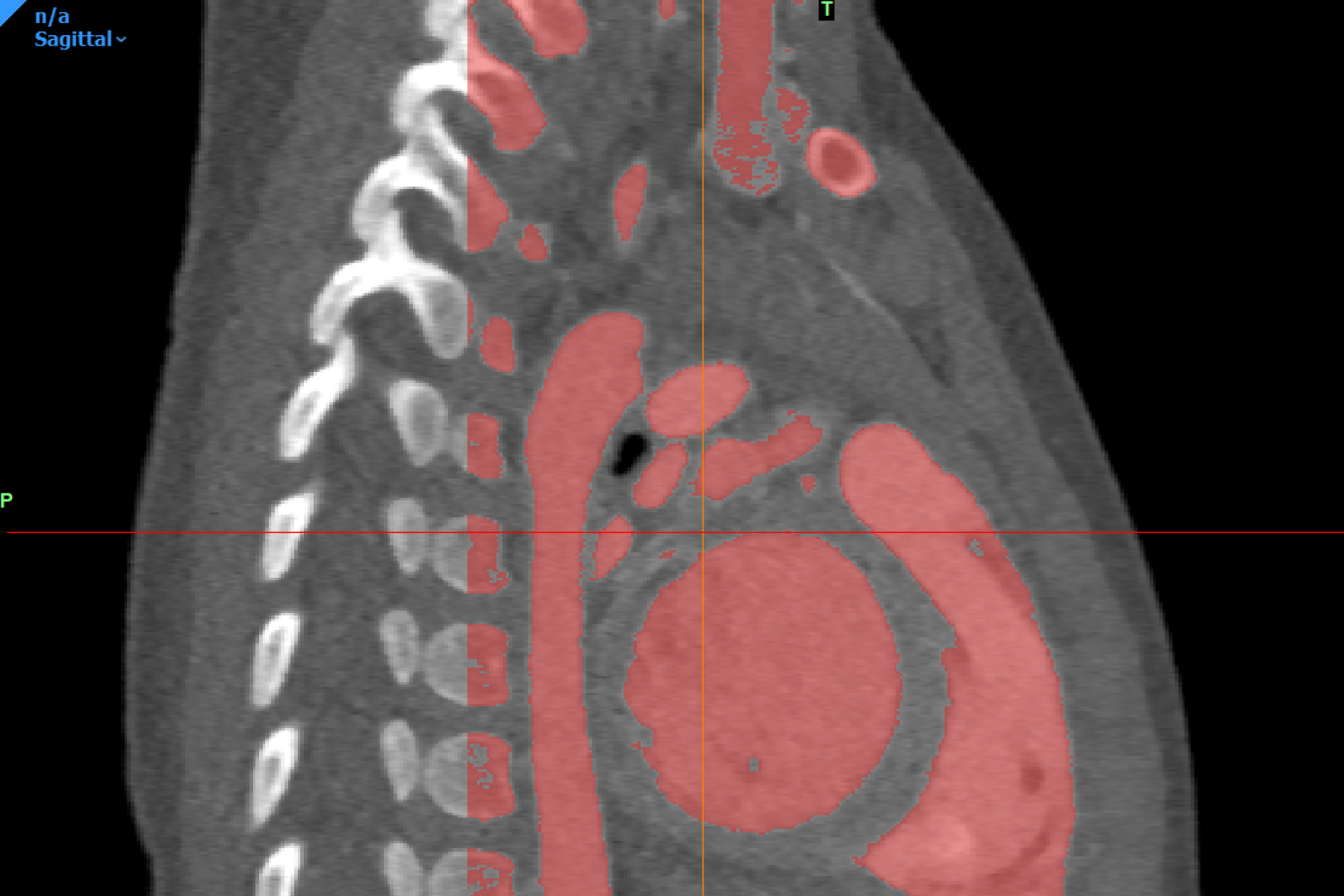
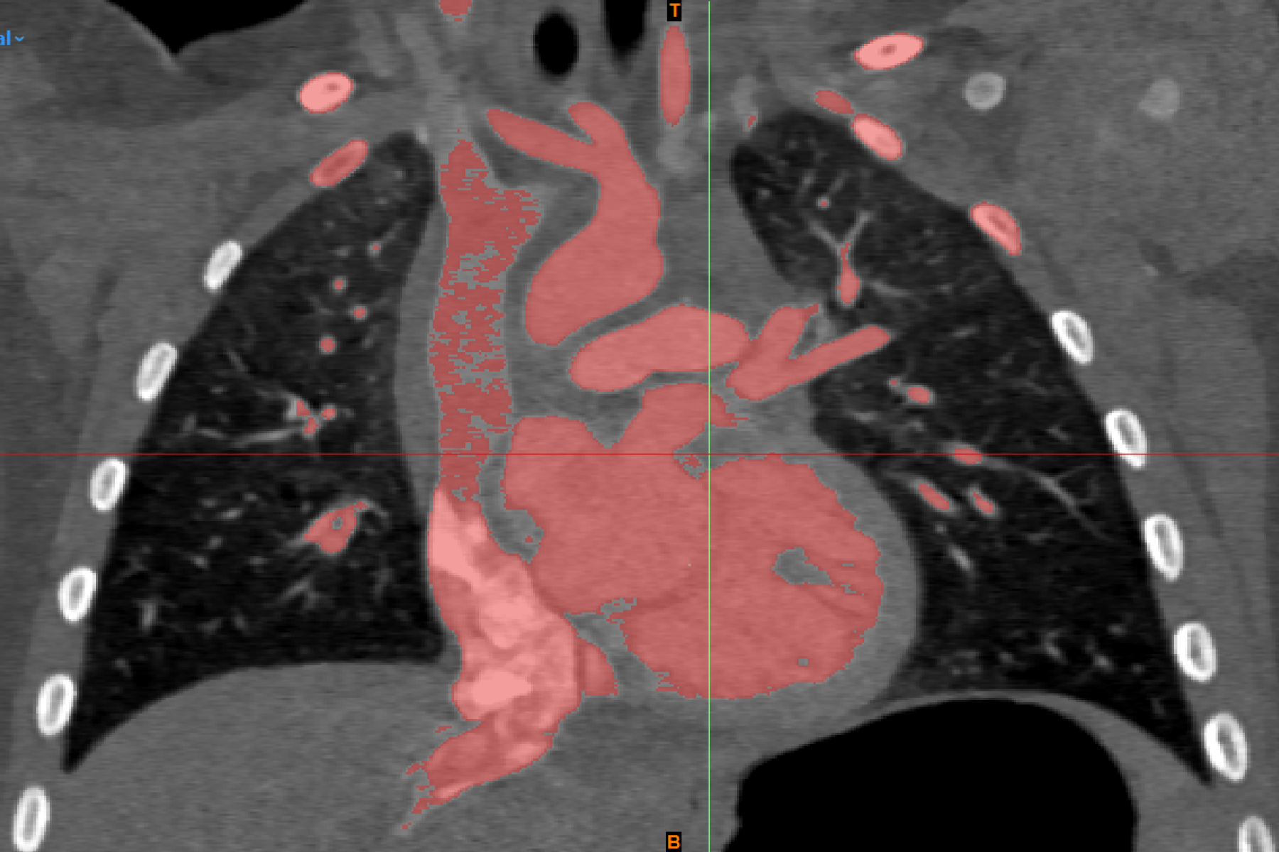
Balloon atrial septostomy is a procedure performed on newborns when the blood vessels leaving the heart are not functioning properly. This procedure is completed with the guidance of ultrasound and provides a temporary solution to improve blood oxygenation of the body until surgery can be performed. Although planning can be done, this surgical procedure is complex, and requires highly skilled fetal cardiologists and a well-prepared catheterization lab. Simulations have become increasingly common to help improve education for surgical trainees. This project aimed to develop a 3D-printed surgical simulator for practicing the atrial septostomy procedure in an effort to enhance clinician learning opportunities, and directly improve the quality of patient care at BC Children’s Hospital.

3D Printing a Heart for Surgical Training
To develop this surgical training tool, the Digital Lab utilized established 3D modeling protocols. The virtual 3D model of the heart was developed with the abnormal anatomical features observed in patients with atrial septostomy, notably, the transposition of the great arteries which impacts the proper flow of oxygenated blood. The model was developed in consultation with a pediatric cardiologist using contrast-enhanced computed tomography scans obtained from infants with this condition. After the virtual model was approved, a physical model was fabricated following standard procedures established by the Digital Lab.

To ensure that the 3D printed heart was able to effectively replicate the elasticity and texture of cardiac tissue and provide a realistic experience to users, flexible and rubber-like materials were utilized during the printing process. Extended blood vessels were included to replicate the access points used by surgeons completing this procedure. A 5mm hole was created in the atrial septum to simulate the atrial septal defect (ASD). This 3D printed heart was then suspended within a simulator developed using Computer-Aided Design (CAD) software. This simulator provides access points that accommodate training for different procedural approaches to ensure that the model is generalizable.
Improving Surgical Education Through Simulation
Currently, the feasibility of the 3D printed simulator is being evaluated by clinicians. To better understand their opinions on this training tool, they complete a post-simulation survey that aims to understand the performance of the model, its realism, and how use of this training tool impacts the users confidence in patient anatomy and surgical technique. The primary goal is to assess the model's effectiveness for balloon atrial septostomy, while the secondary goal is to evaluate its impact on clinician confidence. The anticipated outcome is improved procedural skills and confidence among participants, ultimately enhancing patient care and safety.